Surgical Treatment for Scoliosis
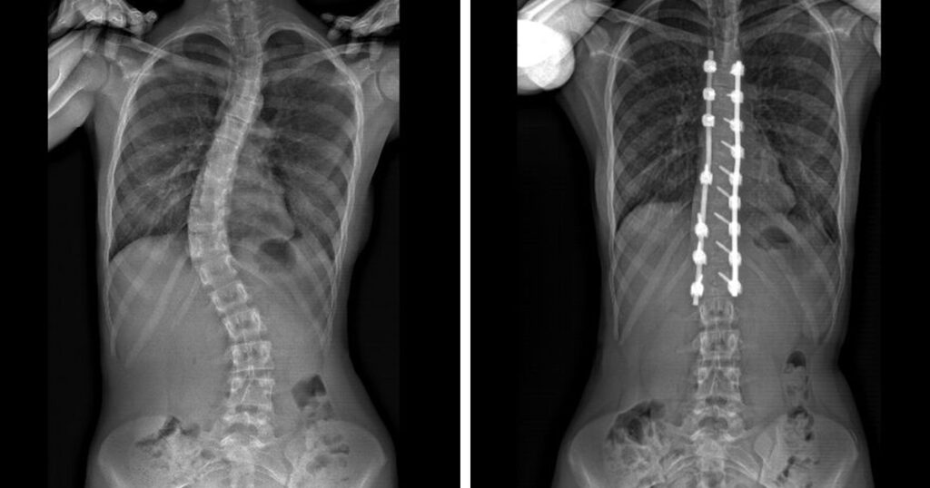
Children with scoliosis may be prescribed exercises to strengthen the back and core, or bracing to correct curvature of a child’s spine over 25 degrees. However, when these are not an option, spinal fusion surgery is the standard of care. This is typically done to correct curves over 40 degrees.
During surgery for scoliosis, rods are inserted and fused to the spine to correct the curvature. Spinal fusion therapy may also be indicated for teenage girls whose curvature is close to but does not exceed the 40-degree mark. A hospital stay would be required.
Rapid Recovery System for Scoliosis Treatment
Today, a new medical technique—the Rapid Recovery System—allows pediatric scoliosis patients to recover more quickly, experience less post-surgical pain and leave the hospital in just a few days.
Developed in 2015 at CHoP in Philadelphia, the Rapid Recovery Joint Replacement program was originally developed to encourage faster recovery (and shorter hospital stays) and reduce pain for patients, as well as expedite treatment and restore function— including getting up and walking within hours after surgery—following joint replacement surgery.
So, what is the Rapid Recovery System? Previously, pain meds (typically opioids) were given to the patient post-operatively as part of a pain management approach. With Rapid Recovery, PRIOR to scoliosis surgery, the patient is given pain medicine such as non-addictive methadone and a steroid after the patient is asleep under anesthesia and just before the surgery begins.
The goal here is to suppress the pain during the surgery so that the child or adolescent never experiences the typical pain stimulus. This in turn prevents the body from releasing chemicals into the body that are usually triggered when there is pain; those chemicals usually cause high blood pressure, muscle contractions and increased pain intensity.
Think of it like this: It is like wetting a house down before a fire.
In other changes to the protocol, physicians previously required their patient to have a bowel movement before resuming eating. However, post-op narcotics tend to slow down one’s bowels. With the Rapid Recovery System, the patient is more likely to experience normal bowel function sooner and eat sooner, which contribute to a faster recovery. And with far less pain, the patient can be up and walking just hours after surgery.
Rapid Recovery at The Pediatric Orthopedic Center
TPOC is the ONLY scoliosis group using the protocol in N.J. Further, we are the only N.J. pediatric orthopedic group that uses custom-made spinal instruments for each patient, which further reduces time in the operating room and speeds recovery.
The Pediatric Orthopedic Center has been offering the Rapid Recovery program to our pediatric scoliosis patients with exceptional results; seven years ago, scoliosis surgery that featured the Rapid Recovery System reduced hospital stays to five days. More recently, the hospital stay for scoliosis surgery has been reduced to just three days, and TPOC, 95% of our pediatric scoliosis patients can return home to their familiar surroundings in just two days.
This revolutionary approach to pain management for scoliosis surgery goes hand in hand with our keen focus on making time to know our patients and their families, and to listen to and address their concerns before the surgery. The pediatric orthopedic surgeon who performs the scoliosis surgery is in the hospital to see patients later that day and the next so that we can stay on top of their progress and answer questions about recovery and post-operative therapies.
Contact TPOC to Discuss the Rapid Recovery Program for Pediatric Scoliosis
Talk to any of our pediatric orthopedic surgeons about the Rapid Recovery System and how your child can recover sooner from scoliosis surgery and with less pain.
How can scoliosis surgery recovery be faster and safer for kids? In this episode, Dr. Rieger from The Pediatric Orthopedic Center discusses the Rapid Recovery Pathway, a treatment approach that helps reduce hospital stays and recovery time for young patients. He explains what families can expect before, during, and after surgery, and how this pathway supports a smoother recovery.


