Foot, Heel and Ankle Conditions
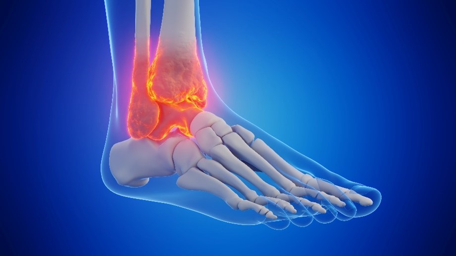
The term “pediatric hindfoot” typically refers to the region of the foot that includes the heel bone (calcaneus) and its connection to the ankle joint.
Pediatric hindfoot conditions encompass various issues impacting a child’s mobility and quality of life, where alignment deformity is a critical concern for diagnosis and management. From overuse injuries prevalent among high-level athletes to trauma-related complications and congenital factors, understanding these conditions is crucial for effective diagnosis and hindfoot treatment.
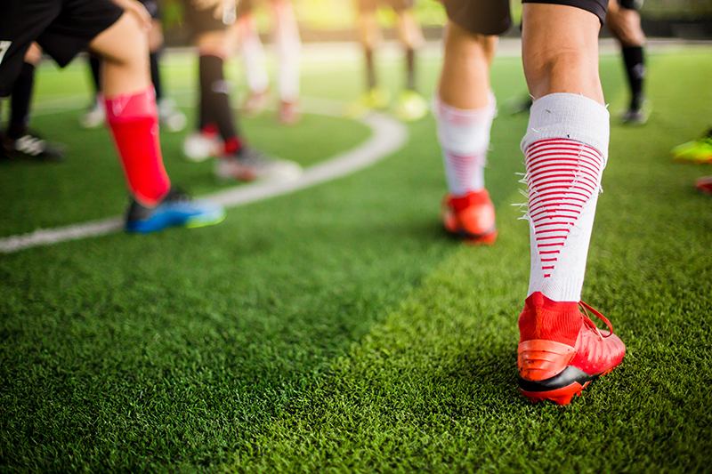
Pediatric Ankle Injuries
Among high-level athletes, hindfoot overuse injuries are prevalent, manifesting as localized inflammation due to strain or excessive use. These injuries, often associated with intense athletic activities, manifest as localized inflammation, particularly in the tendon itself. It’s important to note that this inflammation isn’t prophetic, but rather a response to strain or excessive use.
Trauma from pediatric ankle injuries such as ankle fractures can lead to hindfoot complications, including premature growth plate closure or deformities post-healing. These issues may include premature closure of the tibial growth plate or deformities during the healing process. Additionally, infections or complications post-reductions can contribute to hindfoot problems, necessitating careful monitoring and management.
Deformities and Conditions
The hindfoot can exhibit variations due to syndromes, congenital factors, or a pediatric foot deformity. These variations can differ in their presentation and treatment among various populations due to factors such as genetic predispositions, environmental influences, and healthcare practices in different regions or communities. Post-injury complications like premature growth plate closure or deformities can arise, requiring careful monitoring and management.
Identifying ankle injuries in the context of pediatric hindfoot deformities and conditions is crucial, as it aids in preventing further complications, ensuring accurate diagnosis, tailoring treatment plans, and averting long-term impacts.
Pediatric Hindfoot
Coexisting Conditions and Syndromes: Within the realm of pediatric hindfoot issues, variations often stem from syndromes or congenital factors, presenting diversely and demanding distinct treatment approaches across various populations. The presence of coexisting conditions, such as neuro-muscular issues, congenital anomalies, or prior clubfoot corrections, significantly influences hindfoot development. Understanding these complex associations is pivotal in providing comprehensive care to pediatric patients navigating hindfoot variations.
Commonly Encountered Condition: Inflammation of the calcaneus, commonly known as the heel bone, stands out as a frequent concern within hindfoot issues. This condition often signals ailments such as plantar fasciitis or related problems, particularly prevalent among pediatric patients grappling with hindfoot variations due to syndromes, congenital anomalies, or related factors.
The multifaceted nature of hindfoot presentations and conditions frequently intersects with an array of coexisting factors. These encompass neuro-muscular conditions, congenital anomalies, prior clubfoot corrections, or syndromes specifically impacting hindfoot development. Within this intricate spectrum, inflammation of the calcaneus emerges as one of the most recurrent issues. Notably, it’s potentially linked to plantar fasciitis or analogous concerns, frequently encountered in pediatric patients navigating hindfoot variations associated with syndromes, congenital anomalies, or related influences.
Inflammatory Conditions and Diagnostic Considerations: Exploring the facets of inflammatory conditions affecting the foot, particularly focusing on Achilles tendonitis and its diagnostic considerations, becomes imperative. A crucial aspect highlighted here is the necessity to exclude less common conditions like bone cysts, aneurysms, or stress injuries, which are notably prevalent among young athletes.
Sever’s Disease
Sever’s disease, is a common condition in children experiencing growth spurts. It is a growth plate injury that involves inflammation of the growth plate in the heel bone (calcaneus), typically occurring in active children and adolescents, especially those engaged in activities like running or jumping. The condition arises when the Achilles tendon exerts tension on the heel’s growth plate, leading to pain and discomfort.
Sever’s Disease Treatment
Sever’s disease is not an actual “disease” but rather a temporary condition that tends to resolve once the growth plate fuses and the foot finishes its development. Treatment often involves rest, modifying activities, supportive footwear, and sometimes physical therapy to alleviate symptoms.
Flatfoot
Flatfoot can potentially impact hindfoot issues in pediatric patients due to alterations in the biomechanics and structural support of the foot, including changes such as altered arch support and modifications in gait mechanics.
Flatfoot treatment emphasizes the impact of footwear and home environments, discouraging the use of flexible shoes for better shock absorption and advising against walking barefoot on hard surfaces. Conservative treatments focus on using inserts, supportive shoes, targeted exercises, and physical therapy. Pain management strategies include casting or Cam Walker boots for severe cases, complemented by conditioning and proper positioning during sports activities.
Conditions that Impact the Ankle
Ankle conditions often play a pivotal role in hindfoot issues, interconnecting and influencing the foot’s overall structural integrity and functionality. Among the noteworthy conditions that significantly impact the ankle within the realm of hindfoot concerns are Achilles tendonitis and ankle instability.
Achilles Tendonitis: This condition affects the Achilles tendon, the largest and strongest tendon in the body, connecting the calf muscles to the heel bone. In hindfoot-related complications, Achilles tendonitis can arise due to overuse, sudden increases in physical activity, or biomechanical issues altering the foot’s alignment. It leads to pain, swelling, and stiffness along the back of the heel, potentially affecting hindfoot movements and aggravating existing hindfoot conditions.
Ankle Instability: Ankle instability, characterized by a recurrent “giving way” of the ankle joint, often traces its roots to previous injuries or trauma. This instability can influence the hindfoot by compromising stability during weight-bearing activities, impacting gait patterns, and potentially exacerbating hindfoot-related issues. It’s a critical consideration in hindfoot conditions, as addressing ankle instability becomes integral to ensuring comprehensive care and preventing further complications.
Cerebral Palsy
Cerebral Palsy can affect the hindfoot. It’s a neurological condition that impacts movement and posture, potentially causing muscle imbalance or spasticity, resulting in foot and ankle deformities. Hindfoot valgus, equinus, or planovalgus deformities are frequently linked with Cerebral Palsy, influencing hindfoot alignment and function, thereby affecting an individual’s gait and mobility.
Vertical Talus
Vertical Talus, also known as ‘rocker bottom foot,’ is a congenital foot deformity characterized by an abnormality where the front part of the foot points upward while the heel points downward, creating a convex appearance on the bottom that resembles a rocker. This condition, present at birth, significantly impacts the foot’s structure and alignment, leading to difficulties in weight-bearing, standing, and walking. Without timely intervention, Vertical Talus can result in severe functional limitations and discomfort, requiring specialized orthopedic care to address the structural anomalies and restore mobility.
Coalitions
Coalitions, abnormal bone connections in the foot, play a significant role in hindfoot deformities and intricately affect ankle-related problems. Specifically, conditions like tarsal coalition, where bones fuse abnormally during fetal development, disrupt hindfoot movement, causing discomfort and functional limitations. This fusion often results in stiffness, particularly in the subtalar joint, impacting both hindfoot and ankle mobility, leading to altered gait patterns and misalignments. Diagnosing these complexities typically requires imaging like X-rays or CT scans, and treatment ranges from physical therapy and orthotic devices to surgery in severe cases. Managing conditions like tarsal coalition within ankle and hindfoot issues is vital for comprehensive care, aiming to restore functionality and improve mobility.
Movement-Related Injuries
Achilles Tendonitis
Hindfoot issues in pediatric patients often involve movement-related injuries that significantly impact a child’s mobility and well-being. Among these concerns, Achilles Tendonitis in children stands out as a noteworthy condition affecting kids engaged in active pursuits, such as sports or high-energy physical activities. This inflammatory condition targets the Achilles tendon, the robust connector between the calf muscles and the heel bone, often arising due to overuse, abrupt escalation in physical activity levels, or biomechanical alterations affecting the foot’s alignment.
In the realm of pediatric hindfoot complications, Achilles Tendonitis becomes a prevalent concern due to its potential to disrupt hindfoot movements and exacerbate existing issues. This condition manifests through symptoms like pain, swelling, and stiffness along the back of the heel, impeding a child’s ability to engage in physical activities comfortably. Moreover, in the context of syndromes, congenital anomalies, or other factors influencing hindfoot variations, Achilles Tendonitis poses a heightened risk, potentially complicating the already intricate landscape of foot-related concerns in pediatric patients.
Diagnosing Ankle Injuries
When diagnosing ankle injuries in children within the context of hindfoot complexities, it’s essential to navigate through a range of conditions. This includes distinguishing between more prevalent inflammatory issues like Achilles Tendonitis and rarer hindfoot concerns such as bone cysts, aneurysms, or stress injuries, which, despite their infrequency, can be found in young athletes. Precise diagnosis holds significant importance as it forms the basis for tailored treatment plans. By accurately identifying the specific condition, healthcare providers can devise interventions that aim not only to alleviate symptoms but also to restore hindfoot function, ultimately mitigating potential long-term complications that may affect a child’s mobility and overall well-being.
Non-Surgical Treatments
Conservative treatments stand at the forefront, emphasizing interventions such as inserts, supportive footwear, offloading techniques, and targeted stretching exercises. Physical therapy plays a pivotal role, focusing on specialized stretches for the calf and Achilles tendon, coupled with anti-inflammatory measures. Pain management strategies may involve casting or using Cam Walker boots in severe cases to alleviate pressure and inflammation. Physical therapy remains fundamental, complemented by conditioning and appropriate positioning during various sports seasons to prevent overuse injuries in young athletes. Additionally, less invasive methods, including innovative approaches like the Ponseti or Dobbs techniques, aim to correct deformities and improve foot functionality.
Surgical Treatments
Surgical intervention for pediatric hindfoot ankle injuries is relatively rare compared to non-surgical approaches and is contingent upon factors like the condition’s specifics, severity, and response to conservative treatments. Cases warranting surgery involve scenarios where conservative methods fail to ease symptoms or in the presence of severe deformities, persistent pain, fractures, or ligament damage unresponsive to non-invasive treatments. While these instances exist, they constitute a smaller proportion of pediatric hindfoot ankle injuries compared to those effectively managed without surgery. The decision for surgery is meticulously considered by orthopedic specialists following a comprehensive evaluation, weighing the risks and benefits to ensure the best possible outcome for the child’s functional recovery and overall well-being.
Treating Congenital Defects
Congenital hindfoot defects in pediatric patients often necessitate individualized treatment approaches tailored to the specific defect and its severity. Treatment strategies aim to address functional limitations, restore mobility, and promote proper foot development.
Footwear and Braces
Mild cases of hindfoot issues in pediatric patients can often be effectively managed without surgery. This non-surgical approach involves various methods such as physical therapy, targeted stretching exercises, and the use of orthotic devices or specialized footwear, all aimed at supporting the foot and encouraging proper alignment as the child grows. These interventions, including orthotic devices and suitable footwear, play a crucial role in supporting the foot’s alignment and function as the child’s feet develop. By correcting alignment issues and improving foot function progressively, this comprehensive non-surgical approach aims to guide healthy foot development, often reducing the necessity for more invasive treatments.
The Pediatric Orthopedic Center
If you suspect your child has a foot or ankle injury, contact The Pediatric Orthopedic Center at (973) 538-7700 or by filling out the request an appointment form.
The Pediatric Orthopedic Center is the premier NJ hub for pediatric orthopedics, with four offices throughout northern NJ and two pediatric trained foot and ankle specialists. Having been the leader in pediatric orthopedics in this area for over 25 years, we are the largest and most award-winning pediatric orthopedic practice in the tri-state area.