Approximately 7 in 1,000 children are born with clubfoot in the United States. More children are born with this defect in the right foot than the left, and nearly half of all children with clubfoot are affected in both feet. Boys are twice as likely to be affected by this condition than girls and it affects children of all ethnicities.
What does clubfoot look like? Know the causes and signs
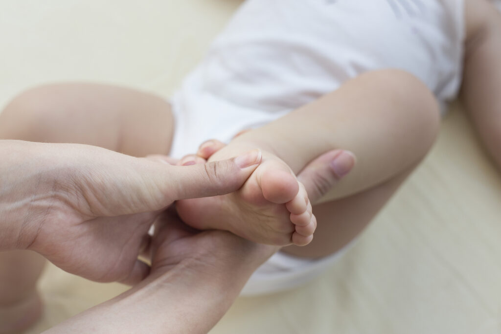
Clubfoot occurs when the tendons and muscles connecting your baby’s heel to the leg are shorter than normal, causing the baby’s foot to turn inward. With clubfoot, a baby’s foot may be up to ½” shorter than the other foot and the calf muscles in the affected leg may be smaller than normal, as well. While not typically associated with other conditions, clubfoot can be found in children with other neuromuscular disorders.
When your child is born, your pediatrician can easily identify clubfoot visually or with X-rays. Clubfoot may also be detected in utero during an ultrasound exam, but parents should know that there is nothing that can be done to correct the condition until birth.
Clubfoot is congenital, with differing opinions about what causes it. Some research suggests a genetic link and if you already have one child with clubfoot, their sibling stands a greater chance of having it, too. Other research suggests that clubfoot is more prevalent in children whose mothers smoked or used recreational drugs while pregnant. Low amniotic fluid levels (oligohydramnios), a viral infection, maternal diabetes, or maternal age have also been associated with the incidence of clubfoot.
While fairly common, the good news is that clubfoot is highly treatable and, in most cases, doesn’t require surgery.
Clubfoot treatment options
While not painful, the treatment of clubfoot should be addressed soon after birth—within a few weeks is ideal. Treatment from clubfoot specialists includes mainly non-surgical and rarely surgical options. Both treatment options have a high rate of success and allow a child to participate normally in life’s activities (walking, sports, dance, etc.).
Until 20 years ago, most treatments for clubfoot involved extensive surgery, including an Achilles tendon tenotomy to lengthen the tendons in the baby’s foot to realign the bones and joints. For the most severe cases of clubfoot, surgery still provides the best assurance that the child’s foot will function normally. However, techniques for the treatment of clubfoot have changed dramatically over the years and in most cases, children don’t require major surgery to correct it.
The good news for most patients is that clubfoot can be successfully treated through orthopedic stretching and reshaping of a baby’s foot. The most accepted and successful non-surgical treatment of clubfoot is the Ponseti method.
Recently, I published a paper with Matthew B. Dobbs, M.D., of The Paley Orthopedic & Spine Institute in West Palm Beach, Florida (a center dedicated exclusively to limb lengthening and the treatment of complex orthopedic conditions) that was just published in the December issue of Clinics in Podiatric Medicine & Surgery that provides a perspective on the history of clubfoot treatment and how the minimally-invasive Ponseti method has become the gold standard of treatment worldwide.
What is the Ponseti method?
The Ponseti method usually begins during the child’s first week after birth when it is the easiest time to manipulate the foot. Since understanding the anatomy of a baby’s foot is so critical to the overall success of this treatment option, it is important to work with a pediatric orthopedist who is certified by Ponseti International to provide this treatment.
The Ponseti method entails stretching the infant’s foot to the correct position and then using a cast to hold it correctly in position. Every week, the pediatric orthopedist will remove the cast to stretch the child’s foot further toward the correct position and place a new cast to hold it in place. This treatment will continue until the child’s foot is facing in the correct direction and usually takes about six weeks.
In some instances, the orthopedist will need to cut the Achilles tendon that connects the child’s heel to the calf muscles so that the tendon can grow to its normal length. This is a brief office procedure done under local anesthetic.
Once the cast is removed, your baby will need to wear specially-designed shoes and a brace for several years at night as the child grows, to maintain correction. In order to prevent a relapse of clubfoot, bracing will be required and usually continues until age 4; this is primarily a nighttime and naptime regimen for much of that time. Parents will also need to do some recommended stretching exercises with their baby once the cast has been removed for a specified period of time.
“Clubfoot is really a misnomer because it is a whole lower limb developmental problem that involves more than just the foot; it involves the child’s leg muscles and bones, as well. As a result, proper treatment requires the attention of a pediatric orthopedist to remedy the condition in the least invasive manner possible,” noted Dr. Dobbs.
Pediatric orthopedists are specially trained to work with children, whose musculoskeletal system is rapidly growing and developing. If your newborn has clubfoot, our team will recommend the appropriate treatment option and long-term protocols to ensure your child has the best possible outcomes for an active life.
If you notice signs of clubfoot, or need a second opinion, please contact us here! If you are looking for our full list of award winning doctors, you can find them here.


