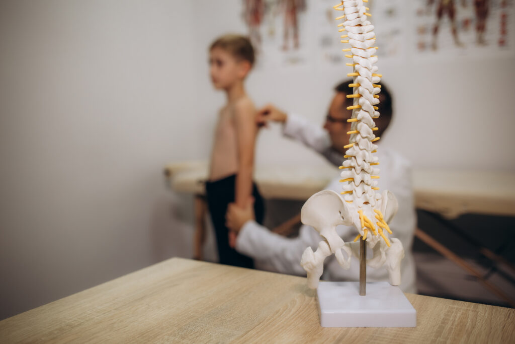
Children and teens with scoliosis often require routine imaging throughout their growth years to monitor changes in spinal curvature and guide treatment decisions. Because scoliosis can progress during periods of rapid growth, having access to accurate imaging is essential for understanding how a child’s spine is developing over time. For many patients, this means undergoing multiple scans throughout their treatment journey, making both precision and safety important considerations.
At The Pediatric Orthopedic Center, advanced technology such as EOS Imaging helps physicians closely monitor scoliosis while prioritizing patient safety. Unlike traditional imaging methods, EOS provides detailed, high quality images with significantly reduced radiation exposure, allowing orthopedic specialists to evaluate spinal alignment, track progression, and make informed treatment recommendations.
Why Regular Scoliosis Monitoring Is Important
Scoliosis is a condition that causes an abnormal curve of the spine, and in children and adolescents, that curve can change as the body grows. Growth spurts, especially during adolescence, are periods when scoliosis progression is most likely to occur. Regular monitoring allows orthopedic specialists to identify whether a curve is remaining stable or becoming more severe.
For children who are diagnosed with scoliosis, consistent imaging provides valuable information that helps guide treatment decisions. Depending on the degree of curvature and the child’s stage of growth, treatment may include observation, physical therapy, bracing, or, in some cases, surgery. Accurate measurements over time help physicians determine the right approach for each patient.
A precise view of the spine is especially important when evaluating scoliosis progression. Imaging allows doctors to compare changes in spinal curvature from one visit to the next and determine whether a treatment plan is working effectively. This ongoing evaluation is a key part of providing personalized pediatric orthopedic care.
The Challenges of Repeated X-Rays During Childhood
Traditional X-rays are a proven tool for evaluating and monitoring scoliosis. Because growing spines often require frequent monitoring, our goal is always to minimize radiation exposure. That is why we prioritize advanced, ultra-low-dose imaging technologies to safely track your child’s spinal development with significantly less radiation. Families often have concerns about the cumulative effects of receiving multiple doses of radiation during childhood. For patients with scoliosis who may need regular imaging from diagnosis through the completion of growth, reducing unnecessary radiation exposure while maintaining accurate imaging is an important goal.
Although traditional X-rays remain an important diagnostic tool, advances in imaging technology have created safer alternatives for patients who require ongoing monitoring. The ability to obtain detailed images while minimizing radiation exposure allows physicians to balance the need for accurate information with a child’s long-term health and safety.
What Is EOS Imaging?
EOS Imaging is an advanced low-dose imaging system designed specifically to provide detailed evaluations of the body’s alignment while reducing radiation exposure compared with traditional imaging methods. Developed by EOS Imaging, the EOSedge system offers 2-D and 3-D imaging capabilities that allow physicians to assess the spine, hips, and other areas of the musculoskeletal system.
Unlike conventional X-rays that typically capture smaller areas of the body, EOS creates a full body image while a patient is standing in a natural, weight-bearing position. This allows physicians to evaluate the relationship between the spine, pelvis, and lower extremities as they function together.
The EOS system captures simultaneous front and side images, creating a more comprehensive, accurate view of skeletal alignment. This provides orthopedic specialists with valuable information that may not be available through traditional imaging alone. While a CT scan can provide detailed images of internal structures, EOS imaging is specifically designed to evaluate full-body alignment with a focus on reducing radiation exposure.
Benefits of EOS Imaging for Children and Teens with Scoliosis
One of the most significant benefits of EOS Imaging for pediatric scoliosis patients is reduced radiation exposure. The EOSedge system uses advanced technology, including Flex Dose™ technology and a photon-counting detector, to adjust radiation levels based on the patient’s size and the area being examined. This allows physicians to obtain high-quality images while limiting unnecessary exposure.
EOS provides weight bearing images, which are especially valuable for scoliosis monitoring because they show how the spine and body are aligned while standing. Since scoliosis affects posture and overall balance, viewing the spine in a natural standing position provides important information for diagnosis and treatment planning.
Another advantage is the quality and detail of the images produced. EOS creates high-resolution 2-D and 3-D images that allow physicians to evaluate spinal alignment, including the relationship between the spine and hips. These detailed images support more accurate measurements and help specialists track even subtle changes over time.
For children and teens, the imaging experience is also designed to be comfortable. The EOS open cabin allows patients to stand naturally during the scan, and the imaging process is completed quickly. This child-friendly approach helps reduce stress while providing the information doctors need.
How EOS Imaging Helps Orthopedic Specialists Track Curve Progression
Monitoring scoliosis requires accurate measurements that can be compared over time. EOS Imaging allows orthopedic specialists to evaluate changes in spinal curvature and overall alignment with detailed, consistent imaging. This information helps physicians determine whether a curve is stable or progressing.
For patients using bracing or other treatments, EOS imaging helps doctors assess whether interventions are effectively controlling curve progression. By providing precise data throughout treatment, EOS supports individualized care plans and helps specialists make informed decisions about the next steps in a child’s treatment.
When Is EOS Imaging Recommended for Scoliosis Patients?
EOS Imaging may be especially beneficial for children and teens who require ongoing scoliosis monitoring. This includes newly diagnosed patients who need a baseline evaluation, children whose curves are being observed over time, and patients receiving active treatment such as bracing.
Because scoliosis treatment often involves monitoring growth and spinal changes over several years, having access to low-dose imaging technology can be particularly valuable. EOS allows physicians to continue tracking progress while reducing radiation exposure during necessary follow-up exams.
What to Expect During an EOS Imaging Appointment
During an EOS scan, the child stands comfortably inside the open cabin while the system captures front and side images of the body. The process is quick, typically taking only a few seconds, and does not require special preparation.
The images produced during the scan provide physicians with a detailed look at the spine, hips, and overall skeletal alignment. Because EOS captures a full-body image in a weight-bearing position, doctors receive information that helps them better understand how the child’s musculoskeletal system is balanced.
Is EOS Imaging Right for Every Child?
While EOS Imaging offers many benefits for pediatric scoliosis monitoring, it is not necessarily required for every patient. Orthopedic specialists determine the most appropriate imaging method based on a child’s age, diagnosis, growth stage, treatment plan, and individual clinical needs.
In some situations, traditional imaging may be appropriate, while other patients may benefit from the detailed information provided by EOS. The goal is always to choose the imaging approach that provides the most useful information while prioritizing patient safety.
Schedule a Consultation with The Pediatric Orthopedic Center
Monitoring scoliosis throughout childhood requires accurate imaging, careful evaluation, and a treatment plan tailored to each child’s needs. At The Pediatric Orthopedic Center, our specialists use advanced technology, including EOS Imaging, to provide precise evaluations while minimizing radiation exposure whenever possible.
If your child has been diagnosed with scoliosis or you have concerns about spinal alignment, our team can help you understand available monitoring and treatment options. Contact The Pediatric Orthopedic Center to learn more about EOS imaging and how advanced technology can support your child’s orthopedic care.